Tuesday 27 January 2015

Abdominal Aorta Ultrasound

I'm rubbish at aorta ultrasounding, and it's one of the few scans that might actually be useful in the ED. I've done my level one course...but it was a while ago, and honestly...I can't remember what I'm looking for anymore. I've written this to try and remind myself...
This is assessed in the ultrasound level one course from CEM, with triggered assessments here.

Anatomy
Xiphoid, through until umbilicus (fourth lumbar vertebra)
- Coeliac axis is 1-2 cm below the diaphragm
- Superior mesenteric artery is 2 cm below the coeliac axis
- Inferior mesenteric artery is 4 cm above the bifurcation
Diameter - <2cm = normal, 2-3cm dilated but not aneurysmal, >3cm aneurysmal


Sens and Spec
Sensitivity of 96.3 % (95 % confidence interval (CI), 81.0 % to 99.9 %)
Specificity of 100 % (95 % CI, 91.8 % to 100 %)
Negative predictive value of 98.6 % (95 % CI, 88.0 % to 99.9 %)
Positive predictive value of 100 % (95 % CI, 86.8 % to 100 %)

Environment
Supine if possible 
Curved probe. 

Technique
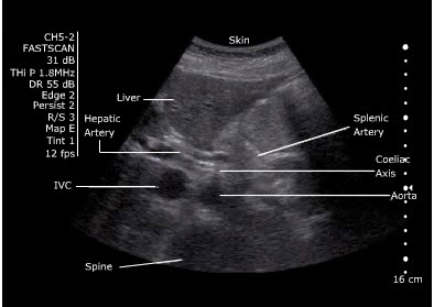
- Start underneath the xiphisternum and identify the aorta  - look for the vertebral body just behind it, liver anteriorly and bowel to the right. 
- Make sure you are not looking at the IVC. 
IVC: Right side, thin walled, compressible, transmitted pulse (‘double bounce’), almond shaped, shape varies
Aorta: left side, thick walled, will not compress, pulsatile, round in shape, constant shape
- Look for the coeliac axis. This is high and often difficult to see. It divides into splenic and hepatic artery - said to resemble the wings of a seagull. This is not essential to find. 


- Sweep to the umbilicus
- Move the probe longitudionally and scan. 
- Look for the "snowman" - where the SMA comes off at the coeliac trunk 
 

Measure
Outer wall to outer wall.
No oblique or angled cuts

Problem Solving- Bowel Gas
“Jiggle” the probe, while applying gentle pressure.  
Reposition the patient. 
Try imaging from below the umbilicus with the probe directed cephalad. 
Try imaging the aortic bifurcation from an oblique angle with the probe placed lateral to the umbilicus (right or left)


References

Thursday 15 January 2015

What did I learn from the ATLS instructor Course?

I had high hopes for the ATLS instructor course, and felt a little under-prepared and didn't really know what to expect. I learnt a lot, and thought I'd share some of my thoughts - in the hope that maybe other people can be better prepared.
Most importantly, ATLS as a course has a really bad reputation. I don't think it's all ATLS's fault - I think that a lot of it is the fault of the individual instructors. Why? Firstly  - slides. You are allowed to omit slides, and add picture slides. You can't alter the text...but there's a lot you can do. Secondly - out of date information - actually, the book may have some of the "older" principles, but the course doesn't focus on them, and allows discussion as long as the principles of ATLS are met.

Preparation
I would suggest:
- Don't read all of the pre-course manual.
- Leave the instructor's manual in its shrink wrap. It's on the CD, and you can read it as a pdf and print off the relevant pages. Open the manual - and then you'll have to decide how to store it in a sensible fashion.
- Re-read the ATLS provider manual. It's there as a pdf on your CD. You need to know it to pass your MCQ - and although you do know it, you need to remind yourself of the "ATLS" way. The MCQs like the random things - like neuroanatomy.

Microteaching
- Plan and rehearse your microteaching.
- The timing is really important - 6 slides, 5 minutes. Title slide, objectives, question and summary = only really 2 slides!
- Interactivity - ask questions
- You can use your own slides or the ATLS slides. Either way make sure you know the slides.

Skill Stations
Prepare by reading the ATLS manual, and watching the DVD. This means you know the "perfect" technique. You don't need to teach the "perfect" technique but you do need to know it -as students will have learnt that. You can then explain why you are deviating from the norm.
You do launch straight into the skills station teaching so make sure it is prepared already.

Critiquing
Stick to Pendleton Plus. It's covered thoroughly in the course, so you should remember it well.

Moulages
You'll be told your moulage scenarios before the course. Take some time to read them. Make sure you bring the moulages + the critiquing form to the moulages.
I struggled a bit to remember everything with the moulages - in retrospect I would highlight the initial scenario (as you need to tell that to the candidate), then write yourself an outline of what happens eg. pneumothorax --> tension --> decompress --> still shocked --> responds to fluids.


Knowledge
There are three causes of error -
 Ignorance --> fixed by lectures and skills
 Ineptitude --> lazyness - fixed by human factors coaching
 Necessary fallibility --> encourage everyone to talk about their errors

Further Reading
- View Sir Ken Robinson's TED talks
- ATLS Manual!

Tuesday 13 January 2015

Exacerbation of COPD

Definition of Exacerbation
You'd have thought that defining an exacerbation would be easy...but everywhere seems to have different exacerbation:
- worsening of the patient's symptoms from stable state that is beyond normal day-to-day variations, and is acute in onset.
- Anthonisen criteria - increased dyspnea, increased sputum volume, and increased sputum purulence 

Investigations
- CXR in all patients coming into hospital 
- ABGs
- ECG
- FBC, U+E

Antibiotics?
- Purulent sputum is often used, but limited accuracy. 
- Bloods - markers of infection are hypothetically useful. 
- GP notebook advises antibiotics if increased in purulent sputum or suspected pneumonia. 

Medical Management
Maximum medical treatment includes:
Controlled oxygen therapy to maintain SaO2 88-92%
Nebulised salbutamol 2.5-5 mg
Nebulised Ipratropium 500 micrograms
Prednisolone 30 mg for 7 to 14 days
Antibiotic agent when indicated
Conflicting evidence about the role of IV magnesium

NIV
NIV should be considered within 60 minutes of arrival to hospital in all patients with an exacerbation of COPD and a persistent respiratory acidosis (pH <7.35 and PaCO2 >6 kPa) in whom medical treatment unsuccessful. 
Patients should improve within four hours - if they don't, consider intubation. 

Exclusion criteria
Life-threatening hypoxaemia
Severe co-morbidity
Confusion/agitation/severe cognitive impairment
Facial burns/trauma/recent facial or upper airway surgery, vomiting, upper airway obstuction, secretions, inability to protect the airway
Haemodynamically unstable requiring
inotropes/pressors (unless in a critical care unit)
Patient moribund
Bowel obstruction
May be used in heart failure or pneumonia

NIV Technique
- Full face mask for 24hours
- Start with an IPAP of 10cm H20, EPAP of 4-5cmH20
- IPAP should be increased by 2–5 cm increments at a rate of approximately 5 cm H2O every 10 minutes
- Bronchodilators, although preferably administered off NIV, should as necessary be entrained between the expiration port and face mask. 

Repeat ABGs:
– after 1 hour of NIV therapy and 1 hour after every subsequent change in settings
– after 4 hours, or earlier in patients who are not improving clinically

Invasive Intubation 
NIV failure or inability to tolerate NIV
Respiratory or cardiac arrest
Respiratory pauses with loss of consciousness or gasping for air
Reduced consciousness or uncontrolled agitation
Massive aspiration
Persistent inability to remove respiratory secretions
Heart rate < 50 with loss of alertness
Haemodynamic instability unresponsive to fluid and vasopressors
Life threatening hypoxaemia

Home or Hospital?

Mortality 
DECAF score

- If intubated and ventilated have an in- hospital mortality of 25%. Two thirds will be dead within a year.  Patients do better than people think. 


References

COPD

There are 30,000 deaths per year attributable to COPD.

Pathogenesis of COPD
 COPD is an umbrella term for any airflow obstruction respiratory term. It includes emphysema, chronic bronchitis, resistent asthma, bronchiectasis and to a certain extent, cystic fibrosis.
Most of it is caused by damage to the lungs from smoking. Cases in young people (younger than 45) should raise the possibility of alpha - 1 - antitrypsin deficiency (2%). Alpha-1-antitypsin protects the alveoli, and deficiency is congenital co-dominant.
Occupational triggers are also possible - heavy exposure to occupational dusts and chemicals, air pollution and cannabis smoking is now being recognised as a cause.


18% of all smokers aged over 35 years will have airflow obstruction
27% of all smokers aged over 35 years with chronic cough will have airflow obstruction
48% of all smokers aged over 60 years and chronic cough will have airflow obstruction

Clinical Presentation
.
Considered in patients > 35 who have a risk factor and one or more of:
exertional breathlessness
chronic cough
regular sputum production
frequent winter "bronchitis"
wheeze


Signs of right heart failure such as raised JVP, peripheral oedema, hepatomegaly


Rule out red flags for other disease before diagnosing COPD (weight loss, effort intolerance, waking at night, ankle swelling, fatigue, occupational hazards, chest pain, haemoptysis)


Investigations
Spirometry:
Airflow obstruction: reduced FEV1/FVC ratio: FEV1/FVC is less than 0.7.
Fixed 70% ratio may mean that COPD is being over-diagnosed in elderly people and under-diagnosed in young people.


Chest X-ray
- Increased bronchovascular markings
- Cardiomegaly
- Lung hyperinflation with flattened hemidiaphragms
- Possible bullous changes.

A full blood count to identify anaemia or polycythaemia
An assessment of body mass index (BMI).


It's difficult to completely differentiate COPD from asthma. NICE guidelines have some recommendations.


Treatment Options
Primary prevention (stopping smoking) is the most important intervention. The next step is to ensure a timely diagnosis. Beta blockers are safe.
Inhalers in COPD are used to prevent and control symptoms, reduce the frequency and severity of exacerbations, improve health status and improve exercise tolerance. Stopping smoking is the only measure that can prevent deterioration.

Tiotropium HandiHaler - long-acting antimuscarinic bronchodilator (LAMA). It is once-daily dosing and the most common side effect is dry mouth.

Seretide - a very expensive inhaler. 


Mucolytics - Increase expectoration of sputum by reducing its viscosity. They can reduce the number of exacerbations and improve symptoms of cough production:
Carbocisteine 750 mg three times daily, reducing to 1.5 g daily in divided doses
Mecysteine 200 mg three times daily for 6 weeks reducing to 200 mg twice daily

LTOT - PaO2 less than 7.3 kPa when stable or a PaO2 greater than 7.3 and less than 8 kPa when stable and one of:
secondary polycythaemia
nocturnal hypoxaemia
peripheral oedema
pulmonary hypertension

End of Life Care
Patients can die quickly after a COPD exacerbation. It is important that their plans for care at the end of life are discussed and documented. Opioids should be used to alleviate breathlessness at the end of life. Benzodiazepines can be considered.

References
See next blog post
Pictures from http://calgaryguide.ucalgary.ca/ 


Tension Pneumothorax


My topic for ATLS instructor teaching is thoracocentesis - so I thought I'd do a little bit of background reading to make sure I can answer all the questions that might come up, admittedly from a "fake" set of learners.

Tension Pneumothorax
- One way valve leak. Air is pushed into the pleural space with no means of escape, so collapses the affected lung.
- The mediastinum is displaced to the opposite side.
- Most common cause is mechanical ventilation.

- Signs include chest pain, air hunger, respiratory distress, tachycardia, hypotension, tracheal deviation AWAY from the side of injury, unilateral absence of breath sounds, elevated hemithorax, neck vein distension, cyanosis.
Classic signs of
tension pneumothorax
Trachea
Expansion
Percussion Note
Breath sounds
Neck veins


Thoracocentesis
5cm needle will reach the pleural space >50% of the time
8 cm needle will reach the pleural space >90% of the time.

Procedure - ATLS Way
- Assess chest and respiratory status
- Administer oxygen and ventilate as necessary
- Identify 2nd intercostal space, midclavicular line
- Surgically prepare the chest. Local anaesthesia if time permits.
- Place patient upright if c-spine injury has been excluded.
- Insert a catheter into the skin, and direct the needle over the rib into the intercostal space.
- Puncture the parietal pleura.
- Remove the needle. Replace the leur lock. Dress.
- Prepare for chest tube insertion

Complications: Local haematoma, pneumothorax,lung laceration, failure
- Placing the needle medially increases the risk of damage to the internal mammary vessels and mediastinum. Lots of experienced ED physicians demonstrated their placement was far too medial.
Emerg Med J 2005;22:788–789


Discussion Points
Cannula - lots of discussion about the length of the cannula. In the UK the cannulas we use seem to be quite short compared to other options. Normal IV cannulae do not reach in up to 65% of cases.
- Some places suggest adding a syringe of saline to the cannula so you can see the bubbles as you go. The cannula can also get easily blocked or kinked.
- CXR first? - recently been called into question. Difficult to know if needle has reached pleura, so a CXR can be helpful. If there is no haemodynamic compromise, wait for a chest x-ray. If there is compromise, do not delay.
Leigh-Smith and Harris recommend urgent CXR first in awake patients, except when:
SpO2< 92% on oxygen
Systolic BP< 90 mmHg
Respiratory rate <10
Decreased level of consciousness on oxygen
Cardiac arrest

- Misdiagnosis?
If you think a patient has a pneumothorax, you decompress the chest, and don't hear  hiss and the patient doesn't improve, and the CXR shows no pneumothorax - do you still need a chest drain? There's no evidence either way - follow local guidelines.

- Tension gastrothorax has similar symptoms. It is caused by a diaphragmatic tear.


References
http://lifeinthefastlane.com/ccc/emergency-thoracocentesis/
http://emj.bmj.com/content/22/11/788.full
http://emj.bmj.com/content/19/2/176.full
http://journal.publications.chestnet.org/article.aspx?articleid=1060258
http://www.trauma.org/archive/thoracic/CHESTtension.html
http://intensivecarenetwork.com/download/emergency-thoracocentesis-doc/
http://emcrit.org/podcasts/needle-finger-thoracostomy/
http://bestbets.org/bets/bet.php?id=783


J Trauma. 2008;64:111–114.
Emerg Med J 2005;22:788–789