Wednesday 28 May 2014

Epistaxis


General
Epistaxis accounts for 1 in 200 visits to the ED. There is a general lack of general first aid knowledge but 85% of patients can be managed without specialist input.
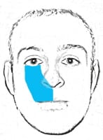
Anatomy
Kiesselbach's plexus = the anastamoses that are joined together. Triangular nasal septum area = Little's area. Most bleeds are from this area (anterior, in 95%). 

Causes
Local minor trauma - nose picking
Drying out in the winter months.
In adults recent alcohol intake, surgery, local malignancy and aneurysm, drugs. 
No studies linking hypertension with epistaxis. 

History Red Flags
    nasal obstruction or congestion
    facial pain
    headaches
    facial numbness, particularly affecting the cheek or side of the nose
    pain around the eye or double vision
    reduced sense of smell
    pain or pressure in one of the ears
In young male patients consider juvenile nasopharyngeal angiofibroma and ask about nasal obstruction, headache, rhinorrhea, and anosmia. These are rare benign tumours that tend to bleed. They occur in the nasopharynx of pre-pubertal and adolescent males.

First Aid Treatment
Remember PPE
Pinch nose (Trotter's Method)
Suck on an ice cube
Ice pack to nose
Ice to neck forehead not shown to help

Further Management


Preparation
- Clean nose with gentle suction. A cut down suction catheter may be less traumatic.
- Might need LA vasoconstrictor applied by a spray or cotton wool pledget. 

- Blood tests not needed unless significant co-morbidity, history or evidence of coagulopathy and disturbance of haemodynamic observatons. Coagulation studies unnecessary unless personal or family history of a coagulation disorder.

- In children, naseptin cream is as good for preventing recurrent epistaxis as silver nitrate but cautery causes more pain. 

Cautery
- Cauterise by direct application for no more than 30seconds in any spot
- If bleeding is too brisk for cautery to be effective cauterise the four quadrants immediately around the bleeding site. 
- Don't do both sides of the nose at once.
- Excess silver nitrate can be removed by application of a saline soaked pledget to the area which neutralises the silver nitrate preventing staining and unwanted burning.

Packs

- All the way in so that you don't get a "Walrus sign". 
- Observe for 30minutes post packing. 
- Observe for longer post pack if:
    Traumatic cause for the epistaxis
    Haemodynamic compromise or shock
    Previous nasal packing within the last 7 days
    Patient is taking anticoagulant medication
    Measured haemoglobin less than 10 g/dl
    Uncontrolled hypertension
    Significant co-morbid illness
    Adverse social circumstances (e.g. the patient lives alone or more than 20 minutes away from the hospital or has no access to telephone or transport)

- Anterior packs for 24 – 48 hours 
- Routine antibiotic cover is not required

- Complications of nasal packing
    Failure to stem bleeding
    Toxic shock syndrome
    Blockage of
        – nasolacrimal duct leading to epiphora
        – sinus drainage leading to acute sinusitis
        – nasal airway leading to hypoxia
    Nasovagal reflex: this reflex occurs during insertion of a pack or instrumentation of the nasal cavity. It leads to vagal stimulation, with consequent hypotension and bradycardia
 
Merocel - easier to insert. 
- Nasal tampons need lubrication with jelly
 
Rapid Rhino - less painful to insert and easier to remove.
- Rapid rhinos need water for at-least 30seconds
 
Foley catheters - advance through nostril until seen in the pharynx. Each balloon should be inflated with 5 - 10mls water and gentle traction applied.

Discharge Advice
Avoid:
    Blowing the nose for one week.
    Sneezing through the nose – keep the mouth open.
    Hot and spicy drinks and food, including alcohol for two days.
    Heavy lifting, straining or bending over.
    Vigorous activities for one week.
    Picking the nose.

References

Sunday 25 May 2014

Dental Blocks

It's on our syllabus that we should be able to do dental nerve blocks - and I suppose we should all be happy to at least give it a try. There are lots of potential dental nerve blocks that can be used. The Oxford Handbook only mentions two of them, so I'm only going to cover those two.

Infraorbital Nerve Block


- Supplies the skin and mucous membrane of the cheek, upper lip, lower eyelid and side of the nose.
- Emerges from the infraorbital foramen (0.5cm below the infraorbital magin and vertically below the pupil).
- Insert the needle into the buccogingival fold between the first and second premolars and direct it up towards the infraorbital foramen.


Mental Nerve Block


- Sensation to the lower lip and the chin
- Emerges from the mental foramen, which is palpable on the mandible on a line between the first and second premolar teeth.
- Block at mental foramen with 1-2ml of LA
- Intraoral or extraoral approach


TMJ Dislocation

Identification
- 90% of cases are bilateral
- Most common cause is excessive mouth opening
- Anterior dislocations are most common.
- Posterior, lateral and superior dislocations are associated with a fracture.


Preparation
- Protect your thumbs with gauze rolls around each thumb.

- Adequate analgesia
- Consider intra-articular lignocaine
- X-ray to confirm reducation and no fracture. May not be necessary if pain settled completely.

Procedure
Massage masseter muscles
Apply rotational force on the mandibular ramus


After Relocation Advice
- Keep jaw closed for next 24hours
- Head bandage if chronic
- Soft diet

References
http://academiclifeinem.com/trick-of-the-trade-massaging-a-mandibular-dislocation-back-in/
http://academiclifeinem.com/trick-of-the-trade-protecting-your-thumbs-in-mandible-relocations/
http://academiclifeinem.com/trick-of-the-trade-stabilizing-mandibular-relocations/
http://crashingpatient.com/medical-surgical/oral-medicine-and-dentistry.htm/
http://academiclifeinem.com/trick-of-the-trade-temperomandibular-tmj-dislocation/
http://www.enlightenme.org/knowledge-bank/cempaedia/mandibular-and-temporomandibular-joint-injuries
http://emedicine.medscape.com/article/149318-overview

Tuesday 20 May 2014

Dental Fractures


The first step is to have a look at the tooth and work out what tooth it is.

Then have a look and try and work out what is wrong with it. See what it looks like. See if it's sore. See if it wobbles.

Concussed
 - no obvious displacement. Tender to touch.
- not wobbly.
 - soft food for a week


Subluxation
- Increased mobility and pain
- Some associated bleeding
- Increased mobility
- Soft food, clean carefully, chlorhexidine mouthwash.

Extrusion
- Partial displacement of the tooth out of its socket
- Partial or total separation of the peridontal ligament resulting in loosening and displacement of the tooth.
- Tooth appears elongated
- If <3mm in an immature developing tooth, needs careful repositioning.

Intrusion
- May or may not intersect the secondary tooth bud.
- May penetrate into the nasal cavity.
- Often associated with alveolar fracture.
- Needs repositioning and careful advice.

Avulsion
- Empty socket
- Do not replace
- Consider x-ray to check not aspirated. Soft food for a week.

In adults
- Wash briefly
- Reposition
- Bite on a hankerchieft to hold it in position
- Glass of milk for storage.
- Flexible splint for two weeks

Infraction
Crack - No follow up needed

Enamel Fracture
Smooth sharp edges

Alveolar Fracture
Manual repositioning + stabilising of the segment
Monitor
Soft diet


Tooth Fractures
These need things done to them. You can catagorise them using the Ellis staging system. I'm going to refer them all to max fax/ a dentist.

Post Extraction Problems

Bleeding - rolled up piece of gauze in the socket for 10min. May need horizontal matress suture - use lidocaine + adrenaline. 

Dry socket pain - if bone exposed. Typically 3- 8 days later. Irrigate with warm saline, oral antibiotics, analgesia and dentist. 


http://www.annemergmed.com/article/S0196-0644%2809%2901141-X/abstract
http://emin5.com/2014/04/07/dental-fractures/
http://www.dentaltraumaguide.org/Permanent_Alveolar_fracture_Description.aspx

Monday 12 May 2014

Dental Emergencies


Incisors grow at 6 - 10months
Canine 16 - 20months
Molars 10 - 24months

Secondary incisors grow at 7 -8 years
Canine + pre-molars at 11- 13years
Molars 6 - 25years

Abscess
Likely to be streptococcus or staph aureus.
In history ask when it started, whether antibiotics used, about presence of systemic features, and immunocompromised. 
Not all patients need antibiotics - give if systemically unwell, high risk or likely complications. 

Amoxicillin or metronidazole - either works.

Admit if systemically unwell, antibiotics no help, rapid spread, dysphagia or dysphonia, immunocompromise or GA needed.

Vincent's Angina
Acute necrotising ulcerative gingivitis or trench mouth
Causes pseudo-membranous infection.
Needs chlorhexidine mouthwash with metronidazole or amoxicillin.
Dental review ASAP. 
 
Ludwig' s Angina - submandibular abscess
Mostly affects males, between 20 -60 years old
Peri-apical abscess of the 2nd or 3rd molar penetrates the inner cortex of the mandible and gains access to the area inferior of mylohyoid. The infection tracks posteriorly so the sublingual space is involved.
The tongue is forced upwards and backwards.
It causes fever, pain, drooling, trismus, dysphagia, submandibular mass and dyspnoea.
Hot potato voice.

Lemierre syndrome
Thrombophlebitis of the jugular veins with distant sepsis of oropharyngeal infection (pharyngitis / tonsilitis +/- peri tonsillar abscess). Caused by an anaerobic gram-negative bacillus.
Patients present unwell, trismus and pain behind the angle of the jaw.

USS shows thrombophlebitis of the internal jugular vein which is often the first hard evidence to suggest Lemierre's.


Wednesday 7 May 2014

Haematology Summary

 I think we've covered everything the syllabus wants us too for adult haematology.


Tuesday 6 May 2014

Acute Leukaemia

As always, check out the Calgary Guide for the pathophysiology of leukaemias. The important thing to realise is that Chronic Myeloid Leukaemia (CML) can progress into AML or ALL.

Chronic Myeloid Leukaemia

Incidence of 1 per 100 000 population. Symptoms are usually chronic and non-specific, but splenomegaly is common and may extend beyond the umbilicus. Lymphadenopathy is not usually prominent. Neutrophilia is common and may be accompanied by thrombocytosis, basophilia, monocytosis, or eosinophilia.


Acute Leukaemias

Acute Lymphoblastic Leukaemia
Rare
Common at 2-10 years with a peak at 3-4 years
Secondary rise after 40 years
Acute lymphoblastic leukaemia is slightly more common among males than females

Acute Myeloid Leukaemia
10-15% of childhood leukaemia but is the commonest leukaemia of adulthood
Incidence increases with age, and the median age at presentation is 60 years.
Acute myeloid leukaemia is equally common among males and females

General
Clinical Features
Bone Failure - signs of anaemia.
  neutropenia - infections of the mouth, throat, skin or perianal region
  thrombocytopenia - spontaneous bruising, menorrhagia, bleeding from venepuncture sites, gingival bleeding or prolonged nose bleeds
Organ infiltration
“B symptoms”- fevers, night sweats, and unexplained weight loss

Investigations
- Anaemia - normocytic
- Low platelets
- Low white cell count - neutropenia with lymphocytosis
- Coagulopathy
- Hyperuricaemia
- Chest radiography is mandatory to exclude the presence of a mediastinal mass

References
http://calgaryguide.ucalgary.ca/slide.aspx?slide=Overview%20of%20blood%20cell%20malignancies.jpg
http://calgaryguide.ucalgary.ca/slide.aspx?slide=Pathophysiology%20behind%20the%20leukemias.jpg

http://www.bmj.com/content/346/bmj.f1660?sso=

Chronic Lymphocytic Leukaemia

There are two types of "chronic" leukaemia. The pathophysiology and clinical features are different - so I'll look at CLL to start with. As always, the Calgary Guide has the best overview. 


Epidemiology
- 3/4 of patients are > 55 years old
- Less than 2% are younger than 45 years old. 
- >20 per 100,000 over 70 years old
- Twice as common in men as women
- Most common in Western White and black populations
- Strongest risk factor is FHx 

Small Lymphocytic Lymphoma
- Same disease, but in the lymph nodes rather than the blood.

Clinical Features
-  80% are asymptomatic
- Painless, often symmetrical lymphadenopathy, splenomegaly, or hepatomegaly. 
- In the latter stages of the disease patients may have anaemia, neutropenia, or thrombocytopenia due to bone marrow failure. 

Investigations
- Raised lymphocytes (lymphocytosis)
- Classified by Rai and Binet systems. 

Treatment
- To slow progression, and for palliation. 

Complications
Infections - bacterial, and viral
Anaemia - bone marrow suppression, or secondary to autoimmune haemolytic anaemia, red cell aplasia, 
Bleeding - bone marrow suppression or immune thrombocytopenia
Transformation- Richter's transformation into CLL. Symptoms of weight loss, fevers, night sweats, muscle wasting, and increasing hepatosplenomegaly and lymphadenopathy. 

References

Friday 2 May 2014

Lymphoma

Haematological malignancies are varied, and there are many different groupings that all present similarly, but with very different clinical prognoses.
14,000 new diagnoses annually.
A full time GP with 2000 patients would see two patients with previously undiagnosed lymphoma every five years. 
Staged with the Ann Arbor system:


Pathophysiology
Lymphoma is a  heterogeneous group of malignancies resulting from abnormal proliferation within lymphoid tissues, such as lymph nodes, spleen, and bone marrow.
They are split into two groups - non hodgkins and hodgkins lymphoma.

NHL
- about 85% - of patients.
- rate of NHL increases with age.
- May be indolent and slow growing or aggressive.

HL
- Reed-Sternberg cells, which have a typical histological appearance. 
- Bi-modal distribution with peaks in young adults and elderly people

Clinical Features

Predisposed to lymphoma:
Immunodeficiency syndromes, EBV, conditions associated with chronic inflammation including H pylori, AU disesases like Hashimoto's, coeliac or Crohn's.

Lymphadenopathy:
        The most common presentation, and can be the only symptom
        Usually presents as a painless lump, often in the neck or supraclavicular region
        Pain can rarely be precipitated by alcohol intake
        The nodes may fluctuate in size spontaneously.
Constitutional symptoms (referred to as B symptoms, present in about 25%):
        Fever (>38°C)
        Night sweats (drenching)
        Weight loss (unexplained, more than 10% of body weight, in the prior six months).
Pruritis

Abdominal masses, especially in children

Investigations
- Full blood count. This may be completely normal. Other pictures include a normochromic, normocytic anaemia, a pancytopenia, a neutrophilia, or an eosinophilia. Anaemia can reflect, for example, chronic disease or marrow infiltration
- C reactive protein and ESR. May be elevated
- Renal function tests. Required to ensure function is normal prior to treatment
- Liver function. May be abnormal in the absence of liver involvement
- LDH. LDH is normally intracellular but is released as a result of cell turnover and cell breakdown due to pressure effects and stress on surrounding tissue
- Uric acid. Some aggressive NHLs are associated with high urate levels that can precipitate renal failure when treatment is started
- Chest x ray. May reveal lymphadenopathy or a mediastinal mass

Hyperviscosity
Haemotological emergency.  The syndrome can arise when there is an elevation in the blood viscosity due to an increase in either red cells, white cells, or plasma components such as immunoglobulins (Igs). This can occur in conditions such as:
    Waldenstrom's macroglobulinaemia (a sub-type of lymphoma) and myeloma (raised monoclonal Igs)
    Polycythaemia (raised red cell volume)
    Acute leukaemias (elevated leukaemia cells in the peripheral blood)
    Myeloproliferative disorders.

Clinical features vary with the underlying condition but can include lethargy, headaches, visual disturbance, isolated cranial nerve palsies, confusion, decreased conscious level, and myocardial infarction and stroke.


References
http://www.enlightenme.org/learning-zone/you-can%E2%80%99t-ignore-paediatric-back-pain
http://calgaryguide.ucalgary.ca/slide.aspx?slide=Clinical%20Features%20to%20Describe%20Abnormal%20Lymph%20Nodes.jpg
http://calgaryguide.ucalgary.ca/slide.aspx?slide=hodgkin%20lymphoma%20-%20pathogenesis%20and%20clinical%20findings.jpg
http://www.enlightenme.org/learning-zone/epistaxis-child
http://learning.bmj.com/learning/modules/flow/ICH.html?execution=e1s1&locale=en_GB&action=start&sessionTimeoutInMin=90&moduleId=10045108&status=LIVE&_flowId=ICH 
http://www.bmj.com/content/348/bmj.g1721?variant=pdf
http://www.enlightenme.org/learning-zone/mass-not-miss

Thursday 1 May 2014

Haemophilia and Bleeding Disorders

von Willebrand's disease - the Calgary Guide

The most common congenital bleeding disorder.
VW Factor and Factor VIII deficiency, and abnormal platelet function
Clinically similar to platelet disorder.
Bleeding is normally mucosal and treated with Factor VIII concentrate

Haemophilia A - from the Calgary Guide


Abnormal Factor VIII which lacks clot-promoting properties
Bleeding in to deep muscles, large joints, or urinary tract.

Desmopressin raises factor VIII levels, and may be sufficient.
Tranexamic acid can help
Otherwise needs factor VIII concentrate to treat

Haemophilia B (Christmas Disease)
Deficiency of Factor XI.
Genetically and clinically indistinguishable from haemophilia A.
Factor IX concentrate to treat
Desmopressin cannot be used in the treatment of haemophilia B.

References
Oxford Handbook of Emergency Medicine
http://www.bmj.com/content/344/bmj.e2707?sso=
calgaryguide.ucalgary.ca/


Warfarin

Warfarin blocks Vitamin K. This reduce the level of clotting factors II, VII, IX and X, the extrinsic clotting pathway.
It prolongs the prothrombin time.This makes people bleed!  The pathophysiology is nicely summarised by the Calgary Guide.


Things that increase INR
- antibiotics, NSAIDs, amiodarone, steroids, thyroxine, phenytoin (unpredictable effect), propranolol,   statins
- liver failure, heart failure, increased metabolic rate - eg thyrotoxicosis, illness, old age

Things that decrease INR- carbamazepine, rifampicin, OCP, vitamin K supplements, barbiturates, herbal remedies
- dietary vitamin K including avocado, leafy green vegetables

General Warfarin Things for Us to Consider
I know we should never have to dose warfarin in the ED...but we do, because we're turning in to a ward. Remember:
    Any change in warfarin dose will have no effect for 16 hours
    Maximum effect is not seen for up to 60 hours
    INR fluctuates - do not change from a stable dose because of one "slightly off" INR
    Small changes in dose can result in large changes in INR - do not overcompensate
    If you change a dose, try to stick with it for at least 2 days before changing it again
    Consider drug interactions, especially antibiotics and steroids
    Any change in the patient’s condition can affect the INR - in particular, worsening liver or cardiac function can dramatically increase the INR
    Only check INR daily if the INR is very high or the patient is clinically unstable

Reversal
- Major bleeding or the requirement for immediate reversal - prothombin complex concentrate and 10 mg IV vitamin K
If giving beriplex, recheck the INR 20minutes later.
Octaplex weight dependent
Beriplex INR dependent

- Minor bleeding - 1-3 mg IV vitamin K
Intravenous (IV) vitamin K works within 6 hours whereas oral vitamin K works completely within 12 hours.
2mg of the IV vit K preparation given PO is the best way to lower an INR and starts to work within six hours

5 or 10mg vit K reverses everything completely.
- INR >8.0 with no bleeding - 1-5 mg oral vitamin K

- INR 5.0-8.0 - warfarin should be withheld unless seen as very high risk of bleeding then vitamin K should be used

Remember, warfarin lasts longer than most clotting factors.


References
http://quizlet.com/27875597/exam-2-ch-41-anticoagulation-drugs-flash-cards/
http://www.enlightenme.org/knowledge-bank/conferences/session/emergency-reversal-anticoagulation-therapy
http://www.doctors.net.uk/ecme/wfrmNewIntro.aspx?moduleid=1539

Low Molecular Weight Heparins

There are lots of LMWHs, and they work on factor Xa and IIa. There's a nice chart on Wikepedia (I know, but it's not important enough to me to chase the link) about the different ratios that they work in. Which is interesting really - I thought they were all pretty similar, but it turns out they are not.


Protamine is used to reverse bleeding from heparin, but also works for the LMWHs.

50mg in 5ml (solution).
Administer by slow IV injection over no greater than 1ml/minute (i.e. 50mg over 5 minutes).
Watch the blood pressure trace closely during administration and slow rate of infusion if the blood pressure drops.
Side effects include anaphylaxis, urticaria and bradycardia.
Protamine provides about 60% neutralisation

Our haematologist said give 1mg per mg of Clexane - another source suggests varying the amount depending on when the Clexane was given. 


References
https://circ.ahajournals.org/content/98/15/1575.full
http://lifeinthefastlane.com/book/critical-care-drugs/protamine/
http://empharmd.blogspot.co.uk/2013/05/protamine-sulfate-for-lmwh.html
http://lifeinthefastlane.com/education/ccc/protamine/
http://journals.lww.com/anesthesiology/pages/articleviewer.aspx?year=1996&issue=11000&article=00037&type=Fulltext

NOACs

There are two main NOACs (Noval Oral Anti-Coagulants) that we are seeing used now. They have limited indications, but these will increase as their trials continue:
- VTE prophylaxis following hip and knee replacement surgery - apixaban, dabigatran and rivaroxaban
- Prevention of stroke and systemic emboli in patients with non-valvular AF - apixaban, dabigatran and rivaroxaban
- Treatment of and secondary prevention of DVT and PE - rivaroxaban 

All of these NOACs work further along the clotting cascade than we are used to, which limits reversal options. 

Dabigatran:
Direct thrombin inhibitor
BD dosing with predictable pharmacokinetics, as no cytochrome p450 interaction. 


Rivaroxaban:  factor Xa inhibitor. Not as much information available - I think similar measures apply. 


Bleeding


   - stop Dabigatran
   - assess severity (if mild just skip a dose)
   - control source of haemorrhage
   - do coagulation screen (APTT, TT)
   - check time of last dose and discuss with Haematology
   - correct co-existant bleeding diathesis e.g. platelets if < 80
   - oral charcoal if ingested within 2 hours
   - haemodialysis (particularly if in renal failure) -> removes ~60% over 2-3 hours

References

http://guidance.nice.org.uk/TA261
http://www.enlightenme.org/knowledge-bank/journal-scan/dabigatran-review-pharmacology-and-management-bleeding-complications-nov
http://emcrit.org/misc/bleeding-patients-on-dabigatran/
http://lifeinthefastlane.com/education/ccc/dabigatran-and-bleeding/
http://emergencyeducation.net/1/category/critical%20care/1.html
http://www.thepoisonreview.com/2011/09/11/dabigatran-toxicity-the-top-10-questions/
http://www.thepoisonreview.com/2011/09/11/3304/
http://i0.wp.com/emcrit.org/wp-content/uploads/Hennepin-County-Dabigatran-Reversal.png

Idiopathic Thrombocytopenic Purpura

ITP is more common in children, than in adults - but I've come across it in the adult syllabus first...so we'll cover kids and adults now.

Pathophysiology
Once again, go no-where else but the Calgary Guide!

- IgG mediated
- Platelets get prematurely destructed. Instead of lasting 8 - 10 days, they only last a few hours.
- Often after a recent cold or other infection
- May be associated with HIV and hepatitis
- May be related to immune disorders or pregnancy.

Epidemiology
- Peak age 2 - 10 years old
- Most cases <5 years
- Can occur in adults in the 3rd and 4th decade
- Acute is more likely to be younger children, and resolves in 1-2 months
- Chronic is >3months, more likely to have underlying disease, rarely remits spontaneously.

Diagnosis
- Platelet count of <100,000 /microLitres
- 40,000 – 90,000 are more concerning for undiagnosed marrow failure or leukemia.
- Normal Hgb and WBC with normal differential.
- The absence of signs of other identifiable causes of thrombocytopenia.

Clinical Features
Cutaneous Bleeding
                Petechiae + Purpura
Mucous Membrane
                Epistaxis
                Wet Purpura – Buccal mucosa, Gingiva, palate, tonsillar pillar purpura or petchiae
                Melena / GIB
                Hematuria
                Menorrhagia
Internal bleeding

Historic points favoring another diagnosis:    Bone/Joint Pain
    Family Hx of easy bruising or low platelets

Exam findings concerning for another diagnosis:    Soft tissue or skeletal morphologic abnormalities
    Nonpetechial rash
    Lymphadenopathy
    Hepatosplenomegaly

Management
Based on clinical findings, rather than absolute platelet count. 
IV 1g + steroids 1mg/kg
no platelets unless life threatening bleed
May need splenectomy

HUS

Haemolytic uremic syndrome is characterised by thrombocytopenia. It is normally triggered by E-coli 0157, which is found in healthy cattle. May also be caused by shigella, yersinia, campylobacter, salmonella
Most cases occur in child
ren <10years, and 2/3 of cases occur in <5years. Incidence is increased if antibiotics or antimotility agents have been used.

Clinical Features
With or without diarrhoea
Diarrhoea may be bloody (70%) or just watery.
Lethargy 2- 14 days after diarrhoea
Neurological symptoms in 33% (irritability, seizures, altered mental status)
Decreased urine output in a patient that is clinically well-hydrated
Pallor
Oedema - often periorbital, in the morning


References
http://pedemmorsels.com/wet-purpura-and-itp/
https://umem.org/educational_pearls/1170/
http://wikem.org/wiki/ITP_in_Pregnancy
http://wikem.org/wiki/Idiopathic_Thrombocytopenic_Purpura
http://www.bestbets.org/bets/bet.php?id=2593
http://www.foamem.com/category/ped-em-morsels/page/11/
https://umem.org/educational_pearls/279/
http://www.guidelinesforme.com/guidelines/133-immune-thrombocytopenia-purpura-itp
http://www.rch.org.au/clinicalguide/guideline_index/Immune_Thrombocytopenic_Purpura/
http://www.pemcincinnati.com/blog/http://bloodjournal.hematologylibrary.org/content/117/16/4190.full
http://www.rch.org.au/clinicalguide/guideline_index/Immune_Thrombocytopenic_Purpura/
http://www.pemcincinnati.com/blog/briefs-itp/http://calgaryguide.ucalgary.ca/slide.aspx?slide=TTP%20HUS.jpg 
http://pedemmorsels.com/nonspecific-diarrheal-illness-or-hus/
http://wikem.org/wiki/Hemolytic_Uremic_Syndrome_%28HUS%29
http://emupdates.com/2009/05/22/916-hemolytic-uremic-syndrome-hus-usual-age-pathophys-ssx-dx-rx/
http://myemergencymedicineblog.blogspot.co.uk/2013/01/what-is-treatment-of-hemolytic-uremic.html
http://wikemerg.ca/wiki/ttphus