Showing posts with label ultrasound. Show all posts

Friday, 26 June 2015

Echo in Life Support

One of the competencies we have to get is echo in life support. I've floated around with echo for a while - but now it's time to knuckle down and be 100% excellent at what we need to do to be level one competent.

- Subxiphoid + one other view, normally parasternal long axis
- Identifies pericardial space
- Look at LV RV (?dilated)
- Identifies IVC, looks at diameter and assesses collapsibility

The Probe
If you are using a cardiac probe, the marker (dot) needs to be on the right to give you the views you are used to. This means that mostly, the probe is near the apex of the heart If you are using an abdominal probe (as many of us have to, especially for the subcostal view) the dot will be on the left.

The Anatomy
Remember that the heart doesn't lie exactly vertically. It lies tilted, and slanted. This helps you to think about what you are seeing.

Subxiphoid or Subcostal View
This is similar to an apical view, but everything is rotated 90 degrees. You can use a curved probe to get this view. Imagine you are shining a torch on the heart. Point it down, and to the patient's left. Remember, the right hand side of the heart is next to the liver.

Because the heart is tilted slightly, the probe hits the liver first, then the right hand side of the heart, and then the left hand side - hence the view you get with the right side of the heart at the top, and the left at the bottom.


Fluid normally collects posteriorly. This view is really good for looking at fluid.

Parasternal Long axis
Aim the probe perpendicular to the chest, just to the left of the sternum in the 4-5th IC space (although I was told that the third is better). The marker should be pointing to the top of the patient. Point towards the patient's right shoulder.

This is really good for looking at LV function, and also shows effusions well.


Remember that the right atrium is likely to be hiding behind the aortic outlet.

Parasternal Short Axis
Rotate the probe to look at the patient's left shoulder, keeping the probe marker on the patient's left. This gives you a view at the bottom of the heart - looking at the ventricles.

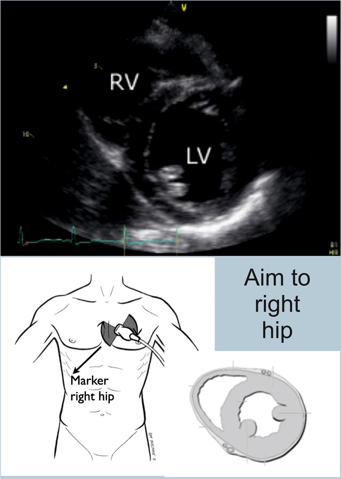
If you scan at the level of the mitral valve, you get the croissant and doughnut appearance. You also get the fish mouth. This is the view we are aiming to get.

Apical Four and Five Chamber Views
Both of these views have the probe in the same sort of position. To get a five chamber view, you point the probe down a little more to be able to see the aorta. The probe marker is on the patient's left.


IVC
Remember that this will be affected by your probe. Start with a subxiphoid view. Rotate the probe vertically, with the probe marker (using a cardiac probe) at the top. Slide 1-2 cm to the right, and tilt towards the heart. This will give you a view of the liver, with the IVC below it, entering the right atrium.
Measure the heart where the hepatic vein is, or 2-3 cm from the heart edge.

Turn into M mode.

The IVC  should collapse on inspiration. If it doesn't, ask the patient to sniff. Measure this as a percentage. If it collapses 25- 50%, they will be fluid tolerant. >50% very likely to be fluid responsive, some say >30%.

I was really struggling to get my head around this anatomy, and one of my collegues drew this brilliant picture for me - and it's mostly clicked. The key I think is thinking about how the heart lies (ie not straight!) and how your probe is looking.


References
http://emergencyultrasound.org.uk/resources/03+Cardiac+views+web.pdf
http://www.rcem.ac.uk/Training-Exams/Training/Ultrasound%20training
http://www.rcem.ac.uk/code/document.asp?ID=5447
https://www.youtube.com/watch?v=oMwgUo6sbyY
https://web.stanford.edu/group/ccm_echocardio/cgi-1bin/mediawiki/index.php/Subcostal_4_chamber_view
http://www.fate-protocol.com/130067GE_Fate_Card.pdf
http://www.smacc.net.au/sonowars-chicago/
http://lifeinthefastlane.com/own-the-echo/

Sunday, 7 June 2015

e-FAST - Pneumothorax

We've talked about FAST...now we need to put the "E" into it. The E wasn't taught to me in my course, and isn't mentioned on the college e-learning module, but it is on their checklist to be assessed for competence...

How
- Ideally you need a linear probe
- Place the probe in the 2nd or 3rd IC space, midclavicular line, and slide caudally.
Bat Sign - the top rib looks like a bat flying out of the screen
- Then look in the 6th IC space, ant axillary line


Normal
Comet Tails - artifacts from the pleural line. Sliding lung and comet tails are normal.
Seashore Sign or waves on a sandy beach- normal lung sliding on M mode.
 - from http://www.intechopen.com/books/hot-topics-in-echocardiography/lung-ultrasound-comet-tails-technique-and-clinical-significance

  - from https://sonospot.wordpress.com/2012/06/19/sonoapp-lung-ultrasound-the-down-low-of-pneumo-with-the-help-of-lichtenstein-of-course/


Pneumothorax
Loss of pleural sliding/ loss of the seashore sign in a spontaneously breathing patient.
This leads to the stratosphere or bar code sign.
No comet tails
 - from https://sonospot.wordpress.com/2012/06/19/sonoapp-lung-ultrasound-the-down-low-of-pneumo-with-the-help-of-lichtenstein-of-course/

References
https://www.acep.org/Clinical---Practice-Management/Focus-On--EFAST---Extended-Focused-Assessment-With-Sonography-for-Trauma/
https://www.youtube.com/watch?v=Yg78aU93SZE
https://www.youtube.com/watch?v=EVQTI7ivhFM
https://sonospot.wordpress.com/2012/06/19/sonoapp-lung-ultrasound-the-down-low-of-pneumo-with-the-help-of-lichtenstein-of-course/

Thursday, 26 February 2015

FAST

I think I make FAST scanning more complicated than it needs to be. Four views - lungs are in eFAST, not FAST...although CEM has them on their checklist.

RUQ - Pouch of Rutherford-Morrison


- Start just anterior to the mid-axillary line, angled slightly backwards.
- Slide the probe towards the head to get a view of the diaphragm
- Look for pleural fluid - a jet black triange just above the diaphragm
- Ask the patient to take a deep breath if possible, particularly if rib shadows obscure the area of interest.

LUQ - Spleno-renal angle
- Posterior-axillary line at about the 6th to 9th intercostal space
- To get rid of rib shadows, and to get a better view of the spleen, slide the probe towarsd the hed and rotate it very slightly clockwise, producing an intercostal oblique view, so that the spleen (not the kidney) is seen.

Pelvic - Pouch of Douglas in females, rectovesical pouch in male
Fluid will collect here.
Place the probe on the pubic bone and note a bone shadow.  Slide slightly towards the head to get a good longitudinal pelvic view.
Full bladder will be triangular in shape.
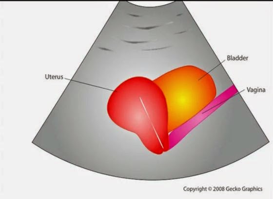
In a female, the body of the uterus sits in the intraperitoneal space just posterior to the bladder (Figure 19), so free fluid will be seen just posterior to the uterus.  This space is often called the pouch of Douglas and sometimes just small amounts can be detected


Cardiac
Subxiphoid view - probe laid almost flat and angled towards the head. Apply probe pressure to indent the epigastrium


Pitfalls
Perinephric fat, especially in obese patients, may be misinterpreted as intraperitoneal free fluid. Consider comparison views between each kidney.
Free fluid isn’t always blood; consider ascites, fluid related to a ruptured ovarian cyst, ruptured bladder or peritoneal dialysis.

90% sensitive and 99% specific for traumatic haemoperitoneum


References
Emergency Ultrasound Made Easy
http://www.trauma.org/index.php/main/article/214/
http://www.sonoguide.com/FAST.html
CEM Website

Tuesday, 27 January 2015

Abdominal Aorta Ultrasound

I'm rubbish at aorta ultrasounding, and it's one of the few scans that might actually be useful in the ED. I've done my level one course...but it was a while ago, and honestly...I can't remember what I'm looking for anymore. I've written this to try and remind myself...
This is assessed in the ultrasound level one course from CEM, with triggered assessments here.

Anatomy
Xiphoid, through until umbilicus (fourth lumbar vertebra)
- Coeliac axis is 1-2 cm below the diaphragm
- Superior mesenteric artery is 2 cm below the coeliac axis
- Inferior mesenteric artery is 4 cm above the bifurcation
Diameter - <2cm = normal, 2-3cm dilated but not aneurysmal, >3cm aneurysmal


Sens and Spec
Sensitivity of 96.3 % (95 % confidence interval (CI), 81.0 % to 99.9 %)
Specificity of 100 % (95 % CI, 91.8 % to 100 %)
Negative predictive value of 98.6 % (95 % CI, 88.0 % to 99.9 %)
Positive predictive value of 100 % (95 % CI, 86.8 % to 100 %)

Environment
Supine if possible 
Curved probe. 

Technique
- Start underneath the xiphisternum and identify the aorta  - look for the vertebral body just behind it, liver anteriorly and bowel to the right. 
- Make sure you are not looking at the IVC. 
IVC: Right side, thin walled, compressible, transmitted pulse (‘double bounce’), almond shaped, shape varies
Aorta: left side, thick walled, will not compress, pulsatile, round in shape, constant shape
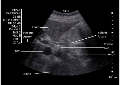
- Look for the coeliac axis. This is high and often difficult to see. It divides into splenic and hepatic artery - said to resemble the wings of a seagull. This is not essential to find. 


- Sweep to the umbilicus
- Move the probe longitudionally and scan. 
- Look for the "snowman" - where the SMA comes off at the coeliac trunk 
 

Measure
Outer wall to outer wall.
No oblique or angled cuts

Problem Solving- Bowel Gas
“Jiggle” the probe, while applying gentle pressure.  
Reposition the patient. 
Try imaging from below the umbilicus with the probe directed cephalad. 
Try imaging the aortic bifurcation from an oblique angle with the probe placed lateral to the umbilicus (right or left)


References