Wednesday, 27 January 2016

Methaemoglobin

Pathogenesis
- Oxidisation of the haem of haemoglobin by free radicals or things like hydrogen peroxide and nitric oxide. 
- Shifts O2 dissociation curve to the left. 

Causes

- Hereditary / Congenital: Hb and NADH-MetHb reductase deficiency
- Acquired: 
  *   Medications eg. Amyl nitrite, Benzocaine, Dapsone, Lidocaine, Nitroglycerin, Nitroprusside, Phenacetin, Phenazopyridine, Prilocaine, Quinones, Sulfonamides (eg. sulfamethoxazole). Chloroquine. 
  *   Chemical agents eg. Aniline dye derivatives (shoe dyes, inks) Butyl nitrite, Chlorobenzene, Nitrate-containing foods, Isobutyl nitrite, Naphthalene, Nitrophenol, Nitrous gases, Silver nitrate, and Trinitrotoluene. Sodium nitrite - used in food preservation. 

Signs & Symptoms

- chocolate brown discoloration of the blood. 
- SaO2 readings go crazy 

0-10% - Features unlikely


10-30%- Mild effects 

Blue-grey ‘apparent’ central cyanosis, fatigue, dizziness, headaches

30-50% - Moderate effects – weakness, tachypnoea, tachycardia


50-70% - Severe effects 

stupor, coma, convulsions, respiratory depression, cardiac arrhythmias, acidosis

> 70% - Potentially fatal


Treatment

<20% - nothing
20 - 30% - oxygen therapy 
>30% - methylene blue
            1-2 mg/kg IV over 5 minutes - 1% (10mg/ml solution) 
            repeat up to 7 mg/kg 
SpO2 normally dives as you give the methylene blue. 
Recheck levels after an hour 

Interesting Note

Hydrogen sulfide poisoning is similar to cyanide poisoning and can be treated by inducing metHb. 


References

http://stemlynsblog.org/feeling-blue-at-st-emlyns/
http://www.rcemlearning.co.uk/modules/papa-smurf-has-a-seizure/
http://emergencymedicineireland.com/2011/07/why-methaemoglobinaemia-is-a-good-thing/
http://emergencymedicineireland.com/2011/07/why-methaemoglobinaemia-is-a-bad-thing/

Sunday, 27 December 2015

Lactic Acidosis

We use lactate a lot, and it is mentioned specifically in the syllabus - it says we should be able to "understand the significance of lactic acidosis in the critically ill patient".

Pathophysiology
Lactate is produced by glycolysis and metabolised by the liver. It is commonly thought it is produced as a result of anaerobic metabolism, but this has been disproved. It is now thought that the adrenergic state and cytokine storm probably change glucose metabolism, changing lactate production.
Adrenaline stimulates beta-2 receptors which up-regulates glycolysis, generating more pyruvate than can be used by the cell’s mitochondria. This excess pyruvate is converted into lactate.  

Lactate can be used by the heart and brain as a fuel source, further suggesting lactate is a stress response.

Lactate clearance is reduced in hepatic failure. The utility of lactate clearance as a marker of disease severity is debated. Some suggest if the lactate level doesn't drop, lactate must still be being produced - so things are serious. The half life of lactate is about twenty minutes.

Measuring Lactate 
The lactate level is independent of anion gap and bicarb so does need to be checked. The level can be high, even if the blood pressure is low.
A venous level is fine, unless there's been a really prolonged tourniquet time when taking the sample!
If you're sending your tests to the lab, even at room temperature (not on ice) the value doesn't change).

Causes of High (>4) lactate
Sepsis
Exercise
Beta-agonist use (like salbutamol)
Even not on ice, the lactate value is unchanged for 15minutes at room temperature.

Treating a High Lactate 
Identify and treat the cause - it's not always sepsis
Remember, a high lactate is a marker of disease severity, not a marker of disease.
Hartmann's (ringers lactate) does not contain the right type of lactate, and does not affect the lactate level.
Remember nebulisers can cause a high latate - which might worsen the dyspnoea.

Causes of a High Lactate
Type A lactic acidosis is seen in states of hypoperfusion or poor oxygenation - shock and cardiac arrest
Type B lactic acidosis is seen in states of high metabolism, organ dysfunction, and toxins. This might be contributed to by thiamine deficiency, neoplastic cells, mitochondrial dysfunction (antiretroviral therapy, linezolid, propofol), hepatic dysfunction.

I haven't written a summary on this one - I think St Emlyns have summarised quite well!!

References
http://emcrit.org/pulmcrit/understanding-lactate-in-sepsis-using-it-to-our-advantage/ 
http://www.emdocs.net/utility-obtaining-lactate-measurement-ed/
http://emj.bmj.com/content/32/9/670.full?hwoasp=authn%3A1451324344%3A4223703%3A1415336879%3A0%3A0%3AVUcQvREASLSBBNQdFnX7Aw%3D%3D
http://emcrit.org/wee/lactate-clearance-flawed/
http://www.heftemcast.co.uk/lactate-lactate-clearance/ 
http://www.ncbi.nlm.nih.gov/pmc/articles/PMC4427204/ 
http://emnerd.com/tag/lactate-clearance/ 
http://emcrit.org/wp-content/uploads/lactate-faq.pdf 
http://www.rcemfoamed.co.uk/portfolio/sepsis-in-the-ed/
http://www.aliem.com/blast-from-the-past-occult-sepsis-lactic-acid-and-mortality/
http://www.oapublishinglondon.com/article/431#
http://emcrit.org/wee/lactate-clearance-flawed/ 
http://www.ncbi.nlm.nih.gov/pubmed/9366769

Tuesday, 28 July 2015

Right Sided Heart Failure

It's often difficult to differentiate right sided heart failure from left sided - because left goes on to cause right!

Pathophysiology

Caused by respiratory problems, and by progression of left sided heart failure. 

Signs
High JVP
Pitting oedema
RUQ pain - hepatomegaly 
Loud P2, and heave 

Investigations
ECG: If the ECG is normal, heart failure is unlikely
  Reduced ECG amplitude (more air between heart and leads)
  Prominent P waves
  Right axis deviation
  RBBB (Broad QRS > 120 ms, RSR’ pattern in V1-3 (‘M-shaped’ QRS complex), Wide, slurred S wave in the lateral leads (I, aVL, V5-6))
  Sagging of ST segment below baseline 
http://lifeinthefastlane.com/ecg-library/basics/p-wave/


P Waves: 
P mitrale (bifid P waves), seen with left atrial enlargement.
P pulmonale (peaked P waves), seen with right atrial enlargement.
P wave inversion, seen with ectopic atrial and junctional rhythms.
Variable P wave morphology, seen in multifocal atrial rhythms.
--> if P waves are inverted check lead placement

Management
Reduce hypoxia and hypercarbia
Ventilation and PEEP
Inotropes and vasopressors
RV assist device may be useful 

http://lifeinthefastlane.com/ecg-library/copd/
http://lifeinthefastlane.com/ecg-library/basics/p-wave/
http://lifeinthefastlane.com/ccc/right-ventricular-failure/
http://www.rcemlearning.co.uk/references/pulmonary-hypertension-and-right-heart-failure/
http://www.rcemfoamed.co.uk/portfolio/rcem-belfast-day-3/ 

Left Heart Failure

Pathophysiology
The left side of the heart fails, for many reasons:
- Restrictive (haemochromatosis, pericardial disease)
- Ischaemic (MI)
- Congestive (volume overload of teventricle from valvular insufficiencies)
- Hypertrophic (longstanding HTN)

This reduces the stroke volume, and hence the cardiac output. 
Cardiac output = stroke volume x heart rate
SV affected by preload, afterload and myocardial contractility. The Frank-Starling Curve has an effect here.

Symptoms
Exertional dyspnoea
Fatigue
PND
Orthopnoea
Vague "not feeling right"

Clinical Findings


- Increased sympathetic activity to attempt to maintain cardiac output = sweating, tachypnoea, tachycardia
- Loud P2, presence of S3 and S4
- Raised diastolic pressure --> narrowed pulse pressure
- Ascites
- Ankle oedema


Investigations
BNP
<100 microgram /mL - unlikely CHF
100 - 500 = may be CHF
500 - most consistent with CHF

CXR


Upper lobe diversion
Peri-bronchial cuffing
Kerley B lines
White interlobular fissures
Pleural effusion
Air bronchogram
Airspace opacification
Cardiomegaly - cardiac ratio > 0.5 on PA film

Echo
Look for evidence of ischaemia and poor contractility

Management Options
Decrease Pre-load: Salt and water restriction
                    Reduce caffeine, alcohol and smoking
                    Diuretics - get euvolemia, then titrate to lowest possible dose.

Improve contractility: Inotropes

Reduce afterload: Vasodilators

Improve Mortality: 
- ACE inhibitors eg. ramipril - NNT 26. Titrate up
- Angiotensin receptor blockers if ACE intolerant.
- Beta blockers eg. metoprolol
- Consider aldosterone agonists like spironolactone NNT 10 or eplerenone NNT 34

End of Life
There is no cure - consider end of life options early. Involve palliative care if needed.

Grading
Class I 
No limitations, ordinary physical activity does not cause undue fatigue, dyspnoea or palpitations (asymptomatic LV dysfunction). 5% 1 year mortality.

Class II 
Slight limitation of physical activity, ordinary physical activity results in fatigue, palpitation, dyspnoea, or angina (Mild CHF). 10% 1 year mortality.

Class III
Marked limitation of physical activity. Less than ordinary physical activity causes symptoms (moderate CHF). 20% 1 year mortality.

Class IV
Unable to carry on any physical activity without discomfort. Symptoms of CHF present at rest (severe CHF). 50% 1 year mortality.


References
http://www.cemfoamed.co.uk/portfolio/agm-conference/
http://learning.bmj.com/learning/module-intro/chronic-heart-failure.html?moduleId=10051928&searchTerm=%E2%80%9Cheart%20failure%E2%80%9D&page=1&locale=en_GB
http://lifeinthefastlane.com/frank-starling-curve-physiology-bscc/
http://calgaryguide.ucalgary.ca/left-heart-failure-pathogenesis/
http://foam4gp.com/2014/10/23/chronic-heart-failure/

Friday, 26 June 2015

Echo in Life Support

One of the competencies we have to get is echo in life support. I've floated around with echo for a while - but now it's time to knuckle down and be 100% excellent at what we need to do to be level one competent.

- Subxiphoid + one other view, normally parasternal long axis
- Identifies pericardial space
- Look at LV RV (?dilated)
- Identifies IVC, looks at diameter and assesses collapsibility

The Probe
If you are using a cardiac probe, the marker (dot) needs to be on the right to give you the views you are used to. This means that mostly, the probe is near the apex of the heart If you are using an abdominal probe (as many of us have to, especially for the subcostal view) the dot will be on the left.

The Anatomy
Remember that the heart doesn't lie exactly vertically. It lies tilted, and slanted. This helps you to think about what you are seeing.

Subxiphoid or Subcostal View
This is similar to an apical view, but everything is rotated 90 degrees. You can use a curved probe to get this view. Imagine you are shining a torch on the heart. Point it down, and to the patient's left. Remember, the right hand side of the heart is next to the liver.

Because the heart is tilted slightly, the probe hits the liver first, then the right hand side of the heart, and then the left hand side - hence the view you get with the right side of the heart at the top, and the left at the bottom.


Fluid normally collects posteriorly. This view is really good for looking at fluid.

Parasternal Long axis
Aim the probe perpendicular to the chest, just to the left of the sternum in the 4-5th IC space (although I was told that the third is better). The marker should be pointing to the top of the patient. Point towards the patient's right shoulder.

This is really good for looking at LV function, and also shows effusions well.


Remember that the right atrium is likely to be hiding behind the aortic outlet.

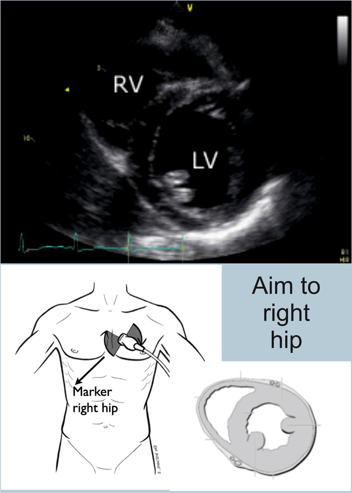
Parasternal Short Axis
Rotate the probe to look at the patient's left shoulder, keeping the probe marker on the patient's left. This gives you a view at the bottom of the heart - looking at the ventricles.


If you scan at the level of the mitral valve, you get the croissant and doughnut appearance. You also get the fish mouth. This is the view we are aiming to get.

Apical Four and Five Chamber Views
Both of these views have the probe in the same sort of position. To get a five chamber view, you point the probe down a little more to be able to see the aorta. The probe marker is on the patient's left.


IVC
Remember that this will be affected by your probe. Start with a subxiphoid view. Rotate the probe vertically, with the probe marker (using a cardiac probe) at the top. Slide 1-2 cm to the right, and tilt towards the heart. This will give you a view of the liver, with the IVC below it, entering the right atrium.
Measure the heart where the hepatic vein is, or 2-3 cm from the heart edge.

Turn into M mode.

The IVC  should collapse on inspiration. If it doesn't, ask the patient to sniff. Measure this as a percentage. If it collapses 25- 50%, they will be fluid tolerant. >50% very likely to be fluid responsive, some say >30%.

I was really struggling to get my head around this anatomy, and one of my collegues drew this brilliant picture for me - and it's mostly clicked. The key I think is thinking about how the heart lies (ie not straight!) and how your probe is looking.


References
http://emergencyultrasound.org.uk/resources/03+Cardiac+views+web.pdf
http://www.rcem.ac.uk/Training-Exams/Training/Ultrasound%20training
http://www.rcem.ac.uk/code/document.asp?ID=5447
https://www.youtube.com/watch?v=oMwgUo6sbyY
https://web.stanford.edu/group/ccm_echocardio/cgi-1bin/mediawiki/index.php/Subcostal_4_chamber_view
http://www.fate-protocol.com/130067GE_Fate_Card.pdf
http://www.smacc.net.au/sonowars-chicago/
http://lifeinthefastlane.com/own-the-echo/

Monday, 8 June 2015

Sore Throat - Tonsillitis

Most sore throats are not caused by a bacterial infection. Make sure there is no epiglottitis or scarlet fever. Scarlet fever would be seen with a rough textured macular rash, with Pastia's lines, and red cheeks with perioral sparing.
There could also be Lemierre's syndrome, retropharyngeal abscess, diptheria, bacterial tracheitis, Ludwig's angina or angioedema.

Red flags for sore throat including significant systemic upset, severe pain, stridor, severe neck stiffness, inability to swallow or tripod position.

Causes
Group A β haemolytic streptococcus - 5-15%
Can cause rheumatic fever in some patients, toxic shock syndrome, necrotising fascitis and post-strep glomerulonephritis. GABHS can be carried asymptomatically - mostly between 3 and 15yrs old (carriage rates 5 - 21%). Adults have much lower carriage rates.
Complications can be suppurative (otitis media, sinusitis, peritonsillar abscess) or non-suppurative (rheumatic fever).
Treat with pen V, 500mg QDS for 10 days.

Viral Tonsillitis
Tonsillitis/pharyngitis are: rhinovirus; coronavirus; adenovirus; herpes simplex; parainfluenza; echovirus; coxsackie A; Epstein-Barr; CMV.

Epstein-Barr Virus
This causes infectious mononucleosis which presents with malaise, headache, fever, pharyngitis, posterior cervical node enlargement, splenomegaly and hepatomegaly. There is a lethargy lasting for six to eight weeks, with suffers being infective for weeks to months. Glandular fever causes anterior and posterior chain lymphadenopathy - bacterial is normally just the upper anterior cervical chain.
Diagnosis is supported by monospot, and there may be atypical lymphocytes and deranged liver enzymes.
90% of patients get an amoxicillin rash with EBV - 5% do with no EBV.

Viral Pharyngitis
Viral infection will often affect immunocompromised and elderly patients. You normally see a unilateral erythema with ulceration. The lesions are often mirrored in the distribution of the glossopharyngeal nerve.

CMV Tonsillitis
This presents very similarly to EBV, and if symptoms persist but the monospot remains negative it should be considered. There is a lot of fever and malaise.

HIV Tonsillitis
Uncommon but should be considered if there is no exudate, tonsillar hypertrophy, rash and mucocutaneous ulceration.

Globus Pharyngeus
This is painless, and patients complain of having "something in the throat", with symptoms improved by swallowing.

Agranulocytosis
Can present with a sore throat, and can be caused by a number of drugs, carbimazole in particular.

Quinsy - peritonsillar abscess
Full, erythematous appearance of the peritonsilar area.
-Deviation of the uvular
-Trismus
- Lateralising pain
- Fullness of the soft palate on the affected side
- Reduction in neck mobility
There is a risk of re-accumulation after drainage. There is no evidence for antibiotics after drainage, but they are generally given.
Quinsy can spread to the parapharyngeal abscess which can be fatal if not treated. Signs can be subtle, and underlying masses might not be fluctuant.

Treatment
Single doses of prednisolone or dexamethasone can be very helpful in major cases - in minor cases they decrease pain by six hours.

Despite their regular use, antibiotics are rarely indicated. Even the SIGN guidelines suggest we use CENTOR guidelines!

The Centor Score - sensitive 97% and specificity 78%
History of fever or temperature > 38oC              +1
Absence of cough                                               +1
Tender anterior cervical lymphadenopathy         +1
Tonsillar swelling or exudates                             +1

Age ≥45 years                                                     -1
A score of 4-5 means that antibiotics should be prescribed.

Antibiotics should be given if there is an increased risk of complications - immuo-suppressed patients, history of valvular heart disease, history of rheumatic fever. If there is an outbreak of GABHS infection within an institution, and a history of repeated episodes of proven GABHS infection.

Streptococcal Score Card - for children 
5 to 15 years
Season (late autumn, winter, early spring)
Fever (≥38.3°C)
Cervical lymphadenopathy
Pharyngeal erythema, oedema, or exudate
No symptoms of a viral upper respiratory infection (conjunctivitis, rhinorrhoea, or cough)

If 5 of the criteria are met, a positive culture for GABHS is predicted in 59% of children; if 6 of the criteria are met, a positive culture is predicted in 75% of children.

Tonsillectomy
Patients with seven attacks in one year, five in two consecutive years, or three attacks in three consecutive years warrant referral to an ENT department.


References
http://www.annemergmed.com/article/S0196-0644(13)00687-2/pdf
http://rebelem.com/patients-strep-throat-need-treated-antibiotics/
http://www.smartem.org/podcasts/treatment-acute-pharyngitis
http://radiopaedia.org/cases/tonsillitis
http://lifeinthefastlane.com/ent-equivocation-002/
http://www.ncbi.nlm.nih.gov/pubmed/?term=Hayward+G%2C+Thompson+MJ%2C+Perera+R%2C+et+al.+Corticosteroids+as+standalone+or+add-on+treatment+for+sore+throat.+Cochrane+Database+Syst+Rev.+2012%3B10%3ACD008268.+http%3A%2F%2Fdx.doi.org%2F10.1002%2F14651858.CD008268.pub2
http://learning.bmj.com/learning/module-intro/sore-throat-diagnostic-picture-tests.html?moduleId=6058292&searchTerm=%E2%80%9Cthroat%E2%80%9D&page=1&locale=en_GB
http://www.sign.ac.uk/guidelines/fulltext/117/index.html
http://www.rcemlearning.co.uk/modules/acute-sore-throat/
http://dontforgetthebubbles.com/tonsillitis/
http://www.rcemlearning.co.uk/references/acute-sore-throat/
http://www.rcemlearning.co.uk/modules/psor-throat/
http://www.rcemlearning.co.uk/modules/sore-throat-with-a-fruity-appearance/

http://www.rcemlearning.co.uk/modules/holiday-sore-throat-are-antibiotics-required/
http://www.aliem.com/modern-em-case-1-and-2-strep-throat/
http://www.aliem.com/paucis-verbis-card-strep-pharyngitis/

Sunday, 7 June 2015

e-FAST - Pneumothorax

We've talked about FAST...now we need to put the "E" into it. The E wasn't taught to me in my course, and isn't mentioned on the college e-learning module, but it is on their checklist to be assessed for competence...

How
- Ideally you need a linear probe
- Place the probe in the 2nd or 3rd IC space, midclavicular line, and slide caudally.
Bat Sign - the top rib looks like a bat flying out of the screen
- Then look in the 6th IC space, ant axillary line


Normal
Comet Tails - artifacts from the pleural line. Sliding lung and comet tails are normal.
Seashore Sign or waves on a sandy beach- normal lung sliding on M mode.
 - from http://www.intechopen.com/books/hot-topics-in-echocardiography/lung-ultrasound-comet-tails-technique-and-clinical-significance

  - from https://sonospot.wordpress.com/2012/06/19/sonoapp-lung-ultrasound-the-down-low-of-pneumo-with-the-help-of-lichtenstein-of-course/


Pneumothorax
Loss of pleural sliding/ loss of the seashore sign in a spontaneously breathing patient.
This leads to the stratosphere or bar code sign.
No comet tails
 - from https://sonospot.wordpress.com/2012/06/19/sonoapp-lung-ultrasound-the-down-low-of-pneumo-with-the-help-of-lichtenstein-of-course/

References
https://www.acep.org/Clinical---Practice-Management/Focus-On--EFAST---Extended-Focused-Assessment-With-Sonography-for-Trauma/
https://www.youtube.com/watch?v=Yg78aU93SZE
https://www.youtube.com/watch?v=EVQTI7ivhFM
https://sonospot.wordpress.com/2012/06/19/sonoapp-lung-ultrasound-the-down-low-of-pneumo-with-the-help-of-lichtenstein-of-course/