Tuesday, 14 April 2015

Myocarditis


There are lots of things that cause myocarditis, but we think it's an immunological thing. It affects children and adults. More likely in immuno-compromised patients - who are more likely to get CMV.

- active viral
   * coxsackie virus B - enterovirus
   * parasitic - Chagas' disease is the most common cause. Endemic in Central and South America
- post viral (lymphocytic - or rheumatic fever)
- hypersensitivity
- autoimmune (SLE, polymyositis, scleroderma, sarcoidosis, DM, thyrotoxicosis, IBD)
- infectious
- giant cell myocarditis
- (HIV is normally another infection rather than being causative)

Symptoms- Chest pain, fatigue, dyspnoea & palpitations
- Often with a prodrome of fever, malaise and arthralgia (89%)
- 60% have a fever, 15% have palpitations
- May be with myopericarditis. 
May be quite hard to differentiate from ACS, especially with troponin rises.

Signs
Fever, tachycardia, pericardial rub + signs of failure. 

Investigations
Cardiac biomarkers may be elevated - but only 1/2 of patients had an elevated troponin 

ECG: non-specific ST segment and T wave changes. Sinus tachycardia. ECG changes not normally in single vessel territory. 
CXR: Normal, or features of cardiomegaly 
Echo: Let ventricular dysfunction in 69% of patients

Treatment
Supportive - inotropes if needed
Immunosuppressive therapy has no evidence.

Prognosis
Heart failure, syncope, BBB or reduced ejection fraction on echo all are associated with poor outcome.


References

Saturday, 4 April 2015

Pulmonary Oedema

Pulmonary oedema - a sign of heart failure, not synonomous

Causes of Pulmonary Oedema
Left ventricle can't empty, causing raised pressure in the lungs. This means the hydrostatic pressure is greater than the oncotic pressure. Fluid moves into the interstitium and alveoli. Some of it is reabsorbed by the lymphatic system. If this can't happen, the accumulating fluid starts to hinder gas exchange, resulting in hypoxia. 
The hypoxia triggers catecholamine release causing vasoconstriction and further left ventricle emptying problems. 

Cardiac Causes: 
Specifically left heart failure and mitral regurgitation

Non Cardiac Causes: NOTCARDIAC or IS NOT THE HEART

I nhaled Toxins (Ammonia, Chlorine, Phosgene, Nitrous oxide, heroin)
S IRS / Sepsis / Septic Shock

N eurogenic (seizure, strangulation, trauma)
O verdose (Heroin, methadone, cocaine)
T hyrotoxicosis

T rauma
H eat (Smoke! Remember to also consider carbon monoxide!)
E lectrocution

H igh altitude pulmonary edema
E mbolism (Pulmonary Embolism, Acute Gas Embolism, Amniotic Fluid Embolism)
A spirin toxicity
R eperfusion or Re-expansion (ARDS)
T ransfusion
asthma, post-obstructive/ post intubation, near drowning

Neurogenic - pulmonary oedema after a significant CNS insult, probably as a result of a catecholamine surg. It's under recognised, and may happen after head injuries. Management is still supportive, and treat the underlying CNS insult.

Presentation
Typically mentioned acute dyspnoea with frothy blood stained sputum. Some of the more recent articles on pulmonary oedema have split the presentation into four categories, and this makes the management and the presentation make a lot more sense.

1. SCAPE  - Sympathetic Crashing Acute Pulmonary (O)Edema)
Often there is no history of CCF, no oedema, no clear ‘trigger’, normal LV function.  This process is largely neuro-hormonal in aetiology and comes on pretty quickly – over hours.

2. Acute-on-chronic (Acute decompensated HF)
Slower onset - over days

3. Acute Causes
These people have a new cause for their oedema - ACS / STEMI, rapid AF / arrhythmia, PE, valve rupture, high output state – sepsis, anaemia. Often a reversible cause.

4. Iatrogenic
Too much IV fluids to treat a chronically low blood pressure!

Clinical Examination
Bibasal inspiratory crepitations or wheeze.
Pale, cold and clammy
Sinus tachycardia or atrial fibrillation
Gallop rhythm may be present.
Murmurs, especially mitral regurgitation and aortic stenosis, may be a precipitating cause.

Assess the JVP, mucus membranes and urine output.
Look for peripheral oedema and hepatomegaly suggesting right heart failure.

Treatment 
- CPAP
Lots of studies suggest the first thing you should do is put CPAP on. BiPAP instead doesn't cause harm - so only use it if there's hypercapnea too. 
Start at 6 cmH20 and rapidly increase to 10cmH20 as tolerated. 

The classic indications for CPAP are: 
pH<7.35
RR >20/min
With "official cautions" being right ventricular failure, cardiogenic shock, severe obstructive airways disease, agitated patient. Contraindications are ET indicated, resp arrest, life threatening hypoxia, unconscious patient.

- Nitrates
Help reduce afterload. 
Resources seem to suggest a higher dose than I've ever used - 400mcg/ minute. As a GTN SL spray is 400mcg/dose, my assumption would be whilst you're mixing up the infusion, get SL in! Put a big bolus in initially to get the GTN up into the therapeutic range. Get an ART line in as soon as possible, so you can control the BP well. 
Be really careful using nitrates in patients with aortic stenosis. 

- Morphine
Morphine is associated with increased mortality, although it makes your patients feel better - so don't give it. They have a theoretical physiological advantage as they reduce pre-load, but this is not born out in real life.

- Diuretics
These have caused much controversy in the NICE guidelines. I've never much liked giving a big dose of furosemide as it takes so long to work - and a lot can happen whilst you're waiting, and it causes a lot of harm. Evidence now suggests that as some patients in pulmonary oedema are euvolaemic, they can make the patients worse.
Diuretics reduced preload (prevent NaCl reabsorption --> increased fluid excretion and vasodilate).

- Inotropes
Commence early, and stop early. Dobutamine -  2-3mcg/kg/min and increase as required.

Investigations
ECG - rarely normal. Often tachycardia, and possibly left ventricular hypertrophy.
 May show precipitating causes - ST segment changes or arrhythmia

CXR -Excludes other causes
cardiogmegaly, interstitial and alveolar oedema and upper lobe diversion.
bat wing or butterfly shadowing - perihilar shadowing
peri-bronchial cuffing
pleural effusions
Kerley lines:
  A lines - course towards hilar. Thickening of the interlobular septa. Only really seen on HRCT
  B lines - 1-2 cm thin lines in the peripheries of the lung. Extend to pleural surface. Perpendicular to the pleural surface. Usually seen at lung bases.
  C lines - short lines that do not reach the pleura
  D lines - B lines seen on lateral CXRs
Differential is diffuse pulmonary haemorrhage - has no dependent gradient, and usually no pleural effusion.

ABG- Type 1 respiratory failure (hypoxaemia)

Bloods 
Take FBC, U&E, LFT, troponin and INR to identify precipitants
Check BNP - produced by stretched cardiomyoctes. No limited identified for acute diagnosis, but BNP is sensitive so can rule out heart failure as a cause of breathlessness - unless the heart failure has happened very very quickly. It can also be raised in sepsis, renal or liver failure, hypoxia, myocardial ischaemia and tachycardia.

Prognosis
Worse outcome if:
Advanced Age
Wide QRS
Low LVF
Hypotension
Precipitated by ischaemia
Previous hospitalisation for heart failure
Marked BNP elevation
Elevated troponin
Hyponatraemia and raised urea and creatinine


References
http://lifeinthefastlane.com/ccc/negative-pressure-pulmonary-oedema/
http://emj.bmj.com/content/22/5/391.1.full
http://emlyceum.com/2011/07/25/acute-pulmonary-edema-answers/
http://boringem.org/2014/07/24/tiny-tips-non-cardiogenic-pulmonary-edema/
http://radiopaedia.org/cases/acute-pulmonary-oedema-1
http://radiopaedia.org/articles/septal-lines-in-lung
http://cdn.lifeinthefastlane.com/wp-content/uploads/2009/11/treatment-of-severe-heart-failu.pdf
http://crashingpatient.com/medical-surgical/cardiology/heart-failure-acute-pulmonary-edema.htm/
http://broomedocs.com/2011/04/clinical-case-004-post-op-peep/
http://broomedocs.com/2011/08/clinical-case-020-the-abc-of-apo/
http://lifeinthefastlane.com/oncology-quandary-004/
http://cdn.lifeinthefastlane.com/wp-content/uploads/2009/11/pulmonary-oedema.pdf
http://lifeinthefastlane.com/aftb-lecture-notes-acute-pulmonary-oedema/
http://lifeinthefastlane.com/ccc/neurogenic-pulmonary-oedema/
http://www.rcemlearning.co.uk/modules/cardiogenic-pulmonary-oedema/
http://www.rcemlearning.co.uk/references/cardiogenic-pulmonary-oedema/
http://lifeinthefastlane.com/ccc/severe-heart-failure-management/
http://radiopaedia.org/articles/pulmonary-oedema
http://www.rcemlearning.co.uk/modules/think-about-it/
http://lifeinthefastlane.com/ebm-acute-pulmonary-oedema/
http://boringem.org/2014/07/24/tiny-tips-non-cardiogenic-pulmonary-edema/
http://www.emdocs.net/furosemide-treatment-acute-pulmonary-edema/?utm_content=bufferc2fc4&utm_medium=social&utm_source=twitter.com&utm_campaign=buffer#comments
http://radiopaedia.org/cases/apo-arrowsjpg
http://radiopaedia.org/articles/bat-wing-pulmonary-opacities-1
http://t.co/sZ7m5DbMTr
http://emtutorials.com/2013/10/killer-sausage/?utm_source=feedburner&utm_medium=email&utm_campaign=Feed%3A+EMTutorials+%28EM+Tutorials%29
http://calgaryguide.ucalgary.ca/Cardiology.aspx
http://lifeinthefastlane.com/book/critical-care-drugs/gtn-ntg/

Thursday, 26 February 2015

FAST

I think I make FAST scanning more complicated than it needs to be. Four views - lungs are in eFAST, not FAST...although CEM has them on their checklist.

RUQ - Pouch of Rutherford-Morrison


- Start just anterior to the mid-axillary line, angled slightly backwards.
- Slide the probe towards the head to get a view of the diaphragm
- Look for pleural fluid - a jet black triange just above the diaphragm
- Ask the patient to take a deep breath if possible, particularly if rib shadows obscure the area of interest.

LUQ - Spleno-renal angle
- Posterior-axillary line at about the 6th to 9th intercostal space
- To get rid of rib shadows, and to get a better view of the spleen, slide the probe towarsd the hed and rotate it very slightly clockwise, producing an intercostal oblique view, so that the spleen (not the kidney) is seen.

Pelvic - Pouch of Douglas in females, rectovesical pouch in male
Fluid will collect here.
Place the probe on the pubic bone and note a bone shadow.  Slide slightly towards the head to get a good longitudinal pelvic view.
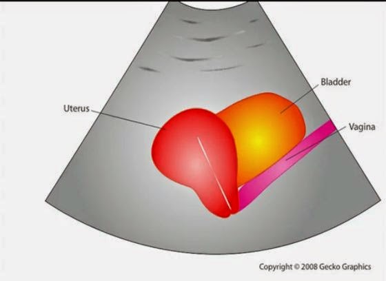
Full bladder will be triangular in shape.
In a female, the body of the uterus sits in the intraperitoneal space just posterior to the bladder (Figure 19), so free fluid will be seen just posterior to the uterus.  This space is often called the pouch of Douglas and sometimes just small amounts can be detected


Cardiac
Subxiphoid view - probe laid almost flat and angled towards the head. Apply probe pressure to indent the epigastrium


Pitfalls
Perinephric fat, especially in obese patients, may be misinterpreted as intraperitoneal free fluid. Consider comparison views between each kidney.
Free fluid isn’t always blood; consider ascites, fluid related to a ruptured ovarian cyst, ruptured bladder or peritoneal dialysis.

90% sensitive and 99% specific for traumatic haemoperitoneum


References
Emergency Ultrasound Made Easy
http://www.trauma.org/index.php/main/article/214/
http://www.sonoguide.com/FAST.html
CEM Website

Monday, 16 February 2015

Carbon Monoxide

I thought I knew about Carbon Monoxide poisoning, and then I went to an (R)CEM day - and learnt a whole lot more about it.

Numbers
4,000 attend ED with CO
200 hospitalised

50 die each year

Pathophysiology

CO binds to haemoglobin with an affinity of approximately 220 times that of oxygen. CO shifts the oxygen dissociation curve to the left. It also binds to myoglobin and mitochondrial cytochrome oxidase, impairing ATP production. Somehow it also binds to platelets so that nitric oxide gets released.

CO binds to fetal haemoglobin and shifts the already left-shifted fetal oxyhaemoglobin dissociation curve further to the left. The half-life of CO in the fetus is longer than in the Mum.
CO is produced continuously in the body as a by-product of haem breakdown. This leads to a normal baseline COHb concentration of about 0.5%. In pregnancy and haemolytic anaemias this can rise towards 5%. 

Animal research – dogs given COHb 65-70%. They died.
2/3 of blood removed, and replaced with already poisoned blood - they were fine. They were then injected with CO, and were also fine. 


- Cigarette smoking leads to COHb concentrations of up to about 12% in heavy smokers 
Non-smokers living away from urban areas have carboxyhaemoglobin concentrations of between 0.4% and 1.0%. 
In an urban or industrial setting, concentrations of up to 5% may be considered normal.

- Methylene chloride (dichloromethane), which is found in some paint strippers and sprays. Once in the liver, is converted to carbon monoxide. Methylene chloride is stored in body tissues and released gradually.

- Electric fires, fires, wood burning stoves, barbeques, shesha pipes gas - anything. 

Clinical Presentation
With prolonged exposure her symptoms could lead to hypotension, seizures, cerebral oedema, metabolic acidosis and respiratory failure.

4.3% of 1758 patients presenting to 4 EDs had raised COHb levels
Unsuspected positive cases 3.5%: 
COPD 7.5% 
Headache 6.3%
Flu-like 4.3% 
Chest pain 3.3% 
Seizures 2.1% 

More Severe Symptoms:
The appearance of intoxication or a personality change
Impaired mini mental-state examination
Vertigo and ataxia
Breathlessness and tachycardia
Chest pain (due to angina or myocardial infarction)
Loss of consciousness 
Seizure or multiple seizures
Abnormal neurological signs including blindness, deafness, and extrapyramidal effects. 
High risk features include chest pain, history of unconsciousness, any continuing neurological symptoms or signs (especially cerebellar features) and pregnancy due to the effects on the foetus

Chronic Presentation
Chronic fatigue
Emotional distress
Memory deficits
Difficulty concentrating
Sleep disturbances
Vertigo 
Neuropathy
Paraesthesias
Recurrent infection
Polycythaemia
Abdominal pain
Diarrhoea

Clinical Assessment

Neuro exam: including tests of coordination and balance, MMSE, short term memory for chronic.

Cherry red skin is only seen in severe poisoning, with levels >20.

Investigations
CO reading taken from a breath analyzer - lactose intolerant patients have raised H2 in their expired breath which can interfere with the readings. 
Venous blood should be taken into anti-coagulant and sent to the laboratory. 

Treatment
Administration of oxygen speeds the elimination of CO from the body. Without therapy, the elimination half life of CO is 4-6 hours. Administration of high flow oxygen by a tight fitting mask at normal atmospheric pressure reduces half life to approximately 76 mins. 
Get your NIV out – you just need the seal and the flow rates of O2 high enough to maintain 100% oxygen. Even manually holding a BVM over them with a good seal does the job.

There is debate about the added value provided by hyperbaric oxygen. A COHb concentration of >20% should be an indication to consider hyperbaric oxygen especially if has any other symptoms:
Loss of consciousness at any stage
Neurological signs other than headache
Myocardial ischaemia/arrhythmia diagnosed by ECG
The patient is pregnant

If metabolic acidosis persists despite correction of hypoxia and adequate fluid resuscitation consider correction with intravenous sodium bicarbonate.


I wrote a powerpoint presentation on this - ages ago - feel free to use and adjust... 


References
Toxbase

Tuesday, 10 February 2015

Cyanide

There was a chemical incident declared once because the pre-hospital team could smell almonds- they thought cyanide could be involved. Turns out someone had been baking... 

Cyanide is released by the incomplete combustion of nitrogen-containing materials, and metabolised from amygdalin. Amygdalin can be found in the pits of many fruits, such as apricots and papayas and raw nuts.

Cyanide is a colourless gas with a bitter almond smell, detectable by only 40% of people.
http://www.christem.com/storage/cyanide%20mechanism
CN blocks cellular respiration by blocking the cytochromes. Affected cells convert to anaerobic metabolism, and a lactic acidosis results. The CNS and the heart are most sensitive.

Symptoms
Symptoms after exposure to high vapor concentrations may include the following:
Transient hyperpnea and hypertension 15 seconds after inhalation
Loss of consciousness in 30 seconds
Respiratory arrest in 3-5 minutes
Bradycardia, hypotension, and cardiac arrest within 5-8 minutes of exposure

Symptoms after exposure to lower vapor concentrations or after ingestion or liquid exposure may include the following:
Feelings of apprehension or anxiety
Vertigo
Nausea, with or without vomiting
Muscular trembling
Loss of consciousness
Headache
Dyspnea

Patients exposed to cyanogen chloride experience severe eye and mucous membrane irritation.Low-dose exposure results in rhinorrhea, bronchorrhea, and lacrimation. Inhalational exposure results in dyspnea, cough, and chest discomfort. Onset of symptoms after exposure to nitriles (acetonitrile and/or propionitrile) may be significantly delayed.

Physical findings of cyanide exposure are generally nonspecific. They classically have cherry red skin.

Laboratory Investigations
- Normal arterial oxygen tension, high venous oxygen tension = decreased AV difference (<10%)
- High anion gap metabolic acidosis
- Raised lactate

Antidotes

Minimal symptoms that resolve spontaneously need observation only.

Other Factors
- Oxygen
- Consider sodium bicarbonate if severe lactic acidosis
- Treat haemodynamic instability and cerebral oedema


References
http://blog.clinicalmonster.com/2015/02/em-ccm-carbon-monoxide-and-cyanide-poisoning/
https://umem.org/educational_pearls/2675/
http://www.enlightenme.org/knowledge-bank/cempaedia/industrial-chemical-incidents

Friday, 6 February 2015

Inhalation

The CEM syllabus says knows the rarer causes of breathless-ness including "inhalational injury from chemical and physical irritants, foreign body inhalation". A search for that came up with cyanide poisoning and methaemoglobinaemia - so have written a bit about them too on further blog posts. I also think that carbon monoxide poisoning is important - blog post to follow.

History
The following key points should be taken in a focused history
            Exactly what exposed to and how much (ie volume and concentration)?
            What route?
            What protective measures were taken?

            What treatment was given?

PPE
Some chemicals do not cause secondary contamination e.g. chlorine.

Smoke Inhalation
- Monitor for 4-6 hours in the ED.
- Observe for longer if high risk items in their history:
History of closed-space exposure for longer than 10 minutes
Carbonaceous sputum production
Arterial PO2 less than 60 mm Hg
Metabolic acidosis
Carboxyhemoglobin levels above 15%
Arteriovenous oxygen difference (on 100% oxygen) greater than 100 mm Hg
Bronchospasm
Odynophagia
Central facial burns
Pulmonary injury from smoke inhalation causes hyperinflation and atelectasis. Debris from cellular necrosis, inflammatory exudate, and shed epithelium combine with carbonaceous material to narrow airways that are already compromised by oedema. Reflex bronchoconstriction further worsens the obstruction.


Metals
Metal fume fever (MFF) is an acute disease induced by intense inhalation of metal oxides- mostly zinc. It is a self-limited syndrome characterized by fever, myalgias, headache, and nausea. Symptoms develop 4-12 hours after exposure and typically last several hours; severe cases generally resolve in 1-2 days. Observation is usually all that is necessary.

The exact pathology of MFF is not well understood but likely involves the deposition of fine metal particulates in the alveoli.


http://www.enlightenme.org/knowledge-bank/cempaedia/industrial-chemical-incidents
http://www.toxlearning.co.uk/course/view.php?id=21
http://emedicine.medscape.com/article/771194-overview
http://emedicine.medscape.com/article/814287-overview
http://emedicine.medscape.com/article/1005903-overview
http://emedicine.medscape.com/article/204178-overview#a0101
http://www.annemergmed.com/article/S0196-0644(05)80424-X/abstract
http://lifeinthefastlane.com/ccc/smoke-inhalation/
http://lifeinthefastlane.com/condom-inhalation/
http://lifeinthefastlane.com/toxicology-conundrum-038/


Tuesday, 27 January 2015

Abdominal Aorta Ultrasound

I'm rubbish at aorta ultrasounding, and it's one of the few scans that might actually be useful in the ED. I've done my level one course...but it was a while ago, and honestly...I can't remember what I'm looking for anymore. I've written this to try and remind myself...
This is assessed in the ultrasound level one course from CEM, with triggered assessments here.

Anatomy
Xiphoid, through until umbilicus (fourth lumbar vertebra)
- Coeliac axis is 1-2 cm below the diaphragm
- Superior mesenteric artery is 2 cm below the coeliac axis
- Inferior mesenteric artery is 4 cm above the bifurcation
Diameter - <2cm = normal, 2-3cm dilated but not aneurysmal, >3cm aneurysmal


Sens and Spec
Sensitivity of 96.3 % (95 % confidence interval (CI), 81.0 % to 99.9 %)
Specificity of 100 % (95 % CI, 91.8 % to 100 %)
Negative predictive value of 98.6 % (95 % CI, 88.0 % to 99.9 %)
Positive predictive value of 100 % (95 % CI, 86.8 % to 100 %)

Environment
Supine if possible 
Curved probe. 

Technique
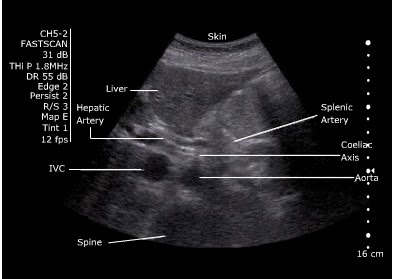
- Start underneath the xiphisternum and identify the aorta  - look for the vertebral body just behind it, liver anteriorly and bowel to the right. 
- Make sure you are not looking at the IVC. 
IVC: Right side, thin walled, compressible, transmitted pulse (‘double bounce’), almond shaped, shape varies
Aorta: left side, thick walled, will not compress, pulsatile, round in shape, constant shape
- Look for the coeliac axis. This is high and often difficult to see. It divides into splenic and hepatic artery - said to resemble the wings of a seagull. This is not essential to find. 


- Sweep to the umbilicus
- Move the probe longitudionally and scan. 
- Look for the "snowman" - where the SMA comes off at the coeliac trunk 
 

Measure
Outer wall to outer wall.
No oblique or angled cuts

Problem Solving- Bowel Gas
“Jiggle” the probe, while applying gentle pressure.  
Reposition the patient. 
Try imaging from below the umbilicus with the probe directed cephalad. 
Try imaging the aortic bifurcation from an oblique angle with the probe placed lateral to the umbilicus (right or left)


References